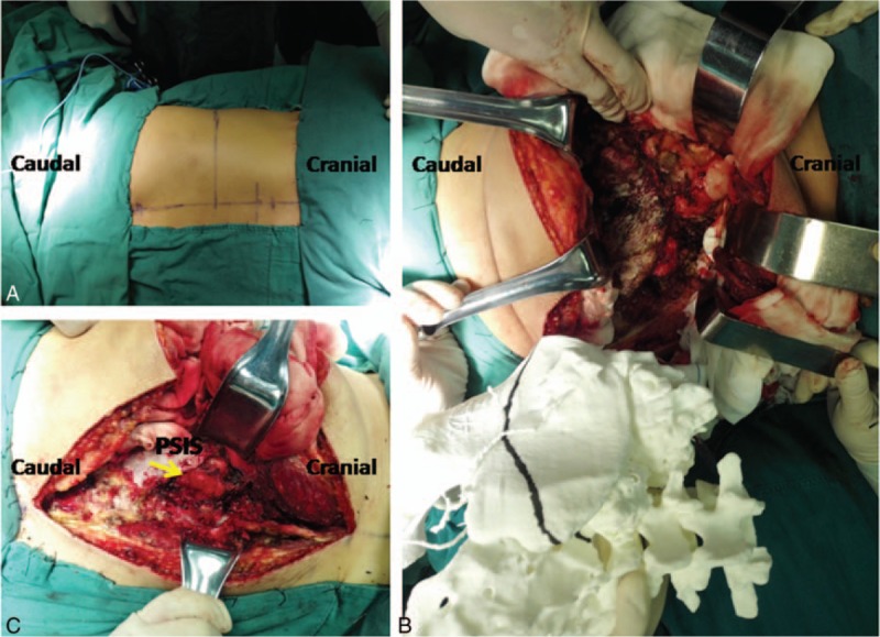
Figure 3.

(A) The incision was converted to a T shape from L4/L5 to the left side. (B, C) Intraoperative photograph showing the use of 3-dimensional model guidance for normal bone removal to expose the tumor (yellow arrow). PSIS = posterior superior iliac spine.
