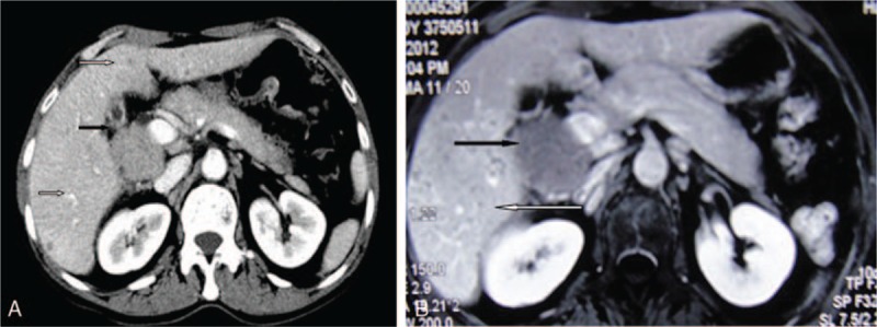
Figure 1.

(A) Abdominal contrast CT scan showed the retroperitoneal mass (black arrow) and the multiple intrahepatic lesions (white arrow). (B) Axial T1-weighted MRI scan showing the retroperitoneal mass with low-intensity image (black arrow) and multiple intrahepatic lesions (white arrow). CT = computed tomography, MRI = magnetic resonance imaging.
