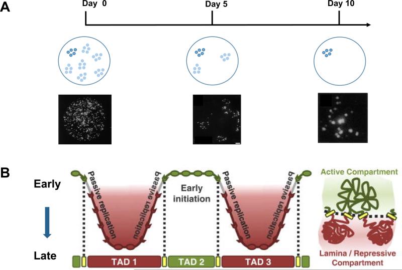
Figure 1. Converging evidence of chromatin domains in animal cells.
A) BrdU pulse-chase labeling followed by immunofluorescence imaging showed that replicon clusters are stable over many cell divisions. A schematic of replication clusters in the nucleus as visualized immediately or 5 days and 10 days after BrdU labeling are shown. The replication clusters remain together even after multiple cell divisions. Image data from Jackson & Pombo (1998). B) Maps of replication domains and chromatin interactions by Repli-chip and Hi-C, respectively, show one-to-one correspondence between replication domains and TADs. A schematic of TADs is on the right. Figure adopted from Pope et al. (2014).

