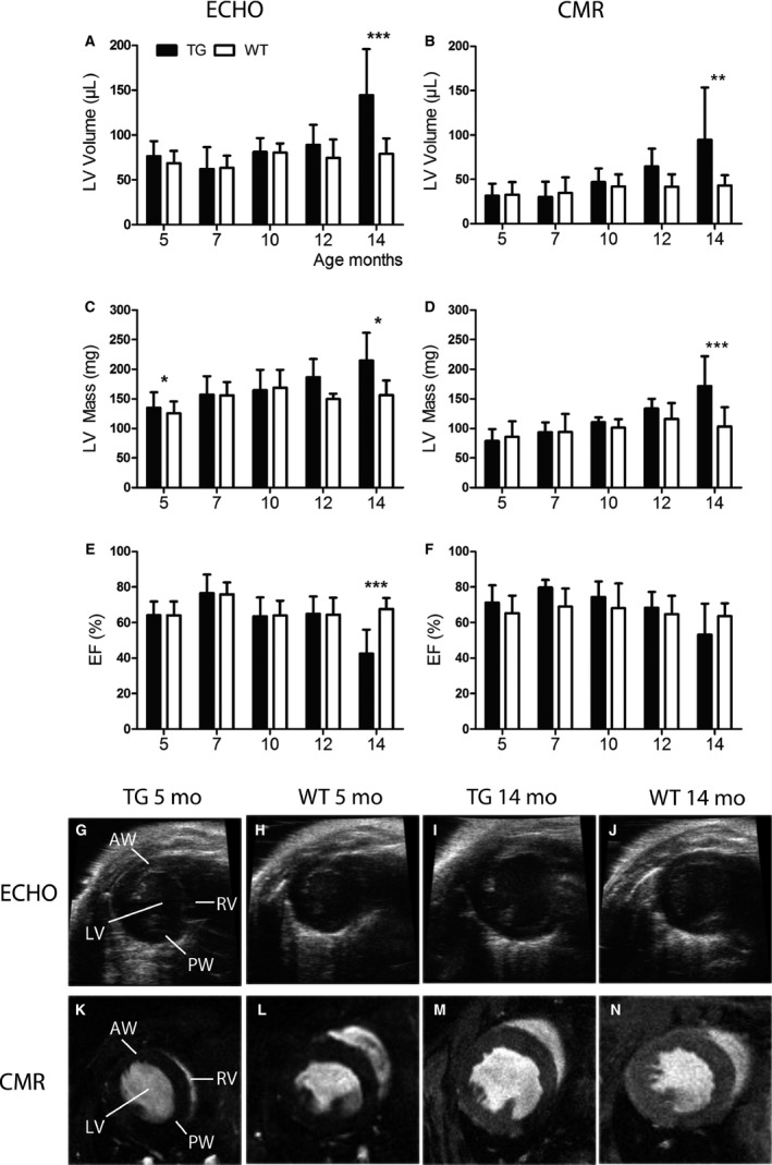
Figure 2.

Measurements of cardiac structure and systolic function by echocardiography and cardiovascular magnetic resonance imaging (CMR). Left ventricular (LV) volume measured by (A) echocardiography and (B) CMR increased slowly with aging in transgenic (TG) mice and a marked dilatation of the LV was observed at the age of 14 months compared to wild‐type (WT) mice. (C, D) LV mass increased with aging in TG mice and at the age of 14 months, the difference compared to WTs was significant measured by both imaging methods. Ejection fraction (EF), measured with (E) echocardiography, decreased significantly in TG mice at the age of 14 months and the trend was similar when imaged with (F) CMR. Representative pictures from TG and WT mice imaged with (G–J) echocardiography and (K–N) CMR at the age of 5 and 14 months. The measurements were done from the mid‐ventricle level at end‐diastole. Mean ± SD, statistical analyses with SPSS linear mixed model analysis. *P < 0.05, **P < 0.01, *** P < 0.001. TG n = 6–11, WT n = 5–10.
