Figure 3.

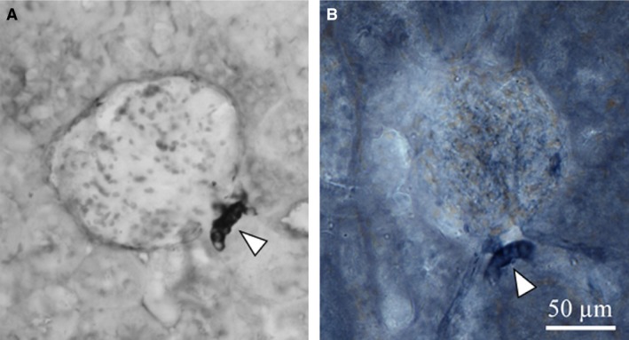
Representative microphotographs of (A) NOS I immunoreactivity (IHC, immunohistochemistry) and (B) NADPH‐d reaction. Arrowheads show the macula densa staining.

Representative microphotographs of (A) NOS I immunoreactivity (IHC, immunohistochemistry) and (B) NADPH‐d reaction. Arrowheads show the macula densa staining.