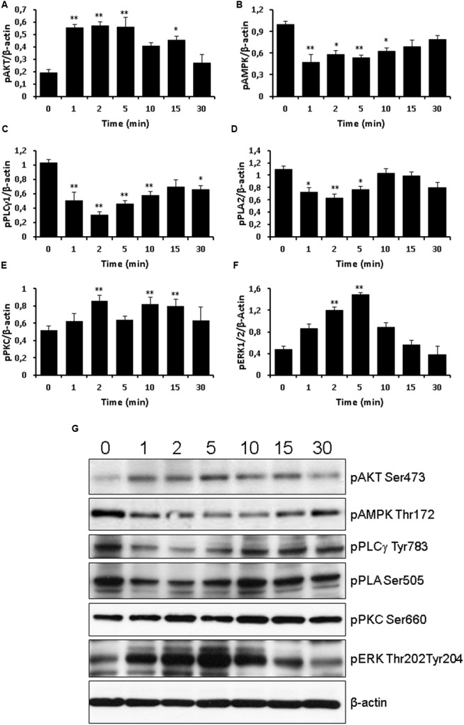
FIGURE 7.

Western blot analysis of signaling phospho-activated proteins. Time course of 10 μM TLQP-21 treatment from 1 min up to 30 min. Equal amounts of cell lysates were processed for Western blot analysis, using specific antibodies for the phosphorylated (active) forms of signaling molecules. Abbreviations: pAMPK = phospho-AMPK (Thr172); pAKT = phospho-AKT (Ser473); pERK1/2 = phospho-ERK1/2 (Thr202/Tyr204); pPKC = phospho-PKC (Ser660); pPLA2 = phospho-PLA2 (Ser505); and pPLCγ1 = phospho-PLCγ1 (Tyr783). To ensure equal loading, blots were also probed with anti-β-actin. (A–F) Results shown are means ± SEM of at least five measurements obtained in five independent experiments. ∗p < 0.05 and ∗∗p < 0.01 vs. time 0. (G) representative images of the blots.
