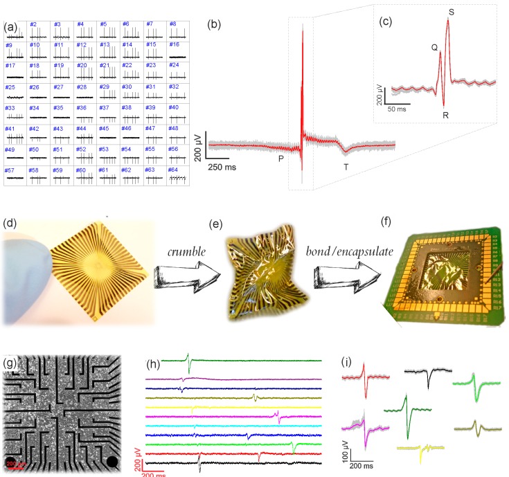
Figure 3.
(a) The spatial resolution map of heart tissue recordings from a GMEA device. The distance between the electrodes is 200 µm in each direction; (b,c) the zoom-in into one action potential of 2 s and 200 ms long are given for a clear observation of P, Q, R, S, and T regions; (d) one flexible chip, which was crumpled (e), then bonded and encapsulated (f); (g) a differential interference contrast (DIC) picture of HL-1 cells grown on top of a GMEA surface; (h) time trace recordings of HL-1 cells from eleven channels on one GMEA chip showing a time delay in recording of different electrodes that reflects spatial propagation; (i) the variety of different HL-1 action potential shapes recorded with the GMEA due to differences in cell–chip coupling.

