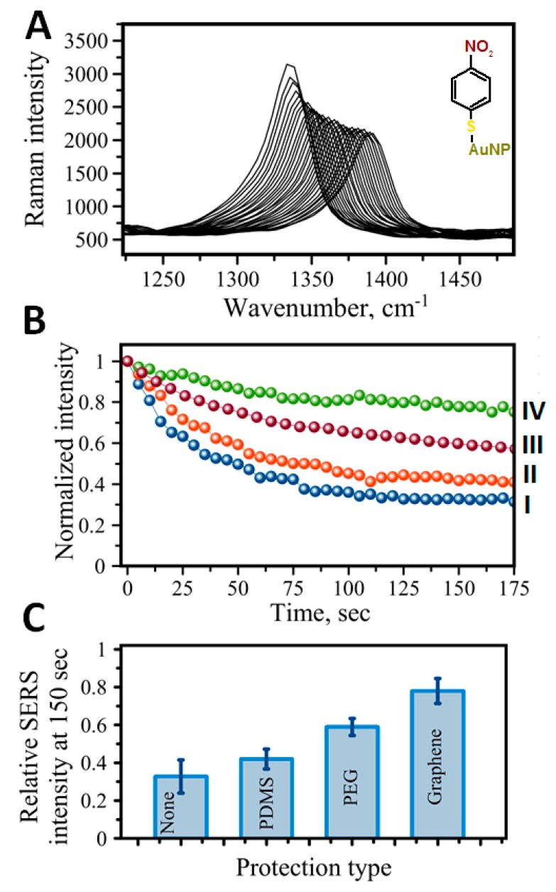
Figure 5.
Protection of SERS-based immunoassay. (A) Time dependence of the signal intensity for the 4-nitrobenzenethiol used as a Raman reporter molecule. Raman peak at 1336 cm−1 corresponding to NO2 symmetric stretch vibration deteriorates with time under laser light exposure (spectra shifted for clarity). (B) A comparative graph showing the intensity of 1336 cm−1 band with time for different protective strategies: (I) no protection, (II) a layer of PDMS, (III) PEG-1000 Da coadsorbed with RRMs on Au nanotag, (IV) graphene monolayer applied on top of immunoassay addresses. (C) Comparison of intensities at 150 s time for protection shown in (B).

