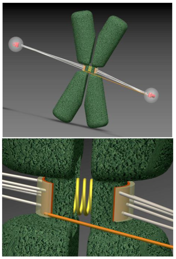
Figure 2.
Merotelic KMT configuration for a congressed chromosome. Top image is a three-dimensional representation of a mammalian chromosome (green) positioned midway between two spindle poles. Sister chromatids (green) are connected by a stretchable centromere, represented as a spring (yellow). Bottom image is an enlargement of sister kinetochores, depicted as semi-transparent layers. Most of the attached MTs are in the proper amphitelic configuration (grey), extending from opposite spindle poles. However, although the chromosome has congressed and its sister kinetochores are positioned back-to-back, they can still bind improper merotelic MTs (one such MT is shown in orange). Computer-generated images are snapshots from video material in [23].

