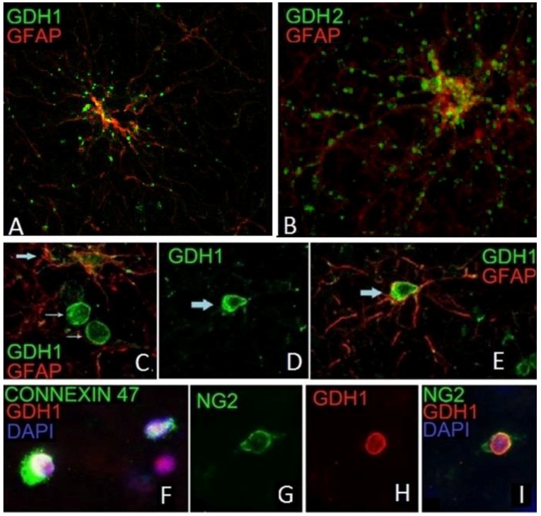
Figure 3.
hGDH1 and hGDH2 expression in glial cells. Punctate immunoreactivity for hGDH1 (A) and hGDH2 (B) is present in the cytoplasm and along the proximal and distal processes of GFAP positive astrocytes (IF images of high magnification). Also, hGDH1-specific staining is detected in the nucleus of a GFAP positive astrocyte (C,D,E), of a connexin 47-positive oligodendrocyte (F) and of an NG2-positive oligodendrocyte-precursor (G,H,I) (IF images of unfixed human frontal lobe cortex. Blue staining represents DAPI labeled nuclei).

