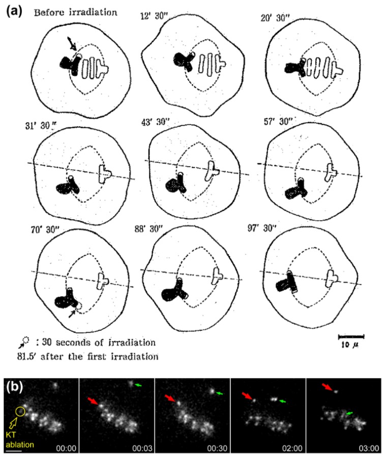
Figure 2.
Evidence that forces on kinetochores are required to position chromosomes at the equator. (a) Original drawings from Izutzu depicting the loss of equatorial position when one of the kinetochore regions from a bivalent chromosome was irradiated with an UV microbeam. Note the displacement of the bivalent from the metaphase plate towards the pole facing the non-irradiated kinetochore after irradiation. Scale bar is 10 μm. Reprinted from Izutsu et al., 1959 [20]; (b) Laser microsurgery of one of the kinetochores from an equatorially-aligned chromosome in a Drosophila S2 cell. Kinetochores were directly labelled with the Centromere Protein A (CENP-A) homologue Cid fused with Green Fluorescent Protein (GFP). Likewise, the chromosome was displaced from the equator after surgery and underwent poleward migration towards the pole facing the undisturbed kinetochore from the pair. Red arrows track the undisturbed kinetochore from the irradiated pair. Green arrows track the congression of an undisturbed chromosome. Laser microsurgery was performed as described in [29]. Scale bar is 2 μm.

