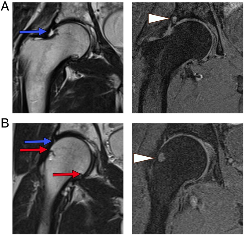
Figure 1.

Typical findings of osteoarthritis at the hip using routine T1-weighted and T2-weighted MRI. (A) Sixty-three years M, T1FS and T2-weighted sequences showing superior joint space narrowing, labral tearing (blue arrow) and an acetabular subchondral cyst (arrow head) adjacent to the labral tear. (B) Fifty-nine years M, T1FS and T2-weighted sequences showing small femoral osteophytes (red arrows), a femoral head–neck junction cyst (arrow head), superior labral tearing (blue arrow) and mild joint space narrowing. M, male; T1FS, T1 fat-suppressed.
