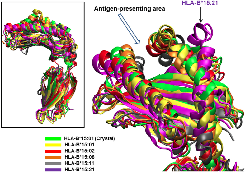
Figure 2. Conformation comparison of HLA-B*15:01 and HLA-B75 protein species.
All simulated structures were aligned with respect to the HLA-B*15:01 crystal structure to visualise the effects due to amino acid changes on protein conformation. The protein conformations at the antigen-presenting area are similar to the HLA-B*15:01 crystal structure (green ribbons), except a larger gap was found in HLA-B*15:21 (purple ribbons with arrow indicated).

