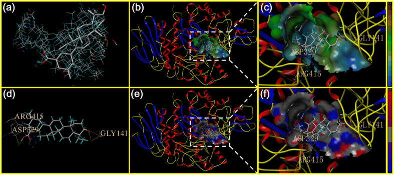
Figure 6. Homology model of the yeast α-glucosidase with analogue UA.
(a) The binding mode of UA docked with the prototype molecular of the active site. (b) and (c) Active site MOLCAD surface representation of lipophilic potential. (d) The active site was surrounded and interacted with the amino acid. (e) and (f) Active site MOLCAD surface representation of hydrogen bonding.

