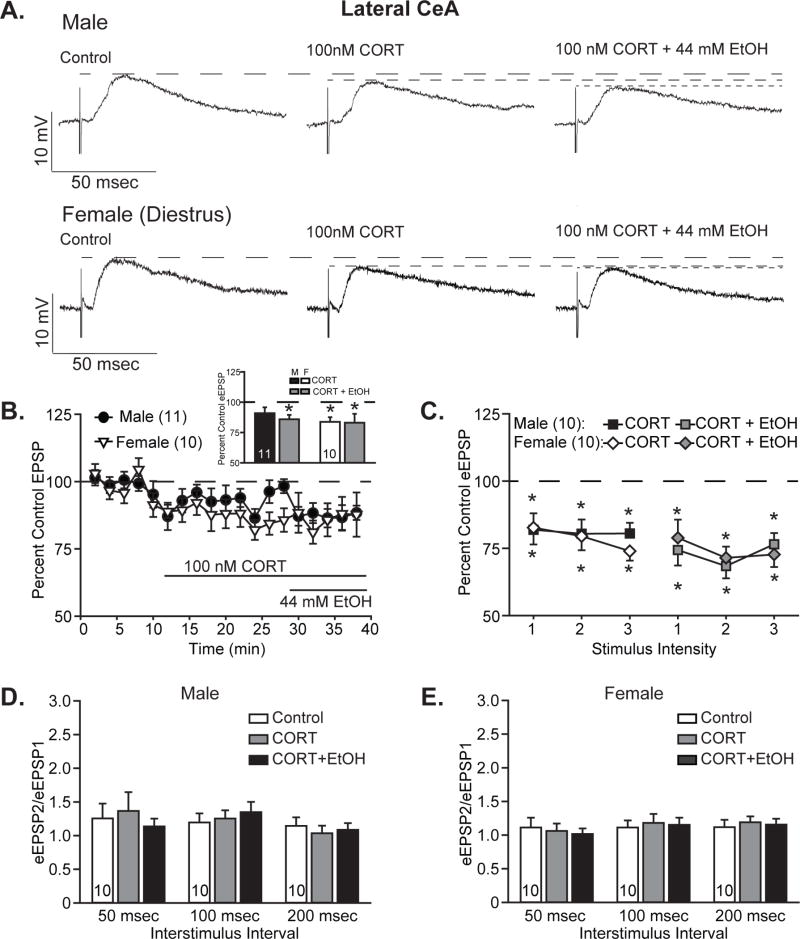
Figure 4. Corticosterone acutely reduced lateral CeA EPSPs and occluded further ethanol effects in females, while males responded more to ethanol.
A. Representative evoked glutamatergic-EPSPs (eEPSPs) in CeL, at baseline (Control) and during superfusion of 100 nM corticosterone (CORT) and subsequent co-application of 44 mM ethanol (EtOH) onto slices obtained from male (top) and diestrus female (bottom) rats. B. Time course of treatment effects, with CORT application following an 8-min baseline and alcohol co-application beginning 20 min after CORT application. Inset shows quantification of the peak CORT- and ethanol-induced changes in eEPSPs over a 4-minute bin beginning not less than 15 min into CORT treatment and not less than 6 min into ethanol co-application. Histograms depict mean ± standard error percent change in eEPSP relative to control. C. Quantification of CORT and ethanol effects on I/O responses to 3 intermediate intensity stimuli in males (squares) vs. diestrus females (diamonds). Data are depicted as mean ± standard error percent change in eEPSP after CORT treatment (black and white shapes) and subsequent ethanol co-application (gray shapes), normalized to control. D–E. Histograms depict mean ± standard error paired pulse ratios for male (D) and female (E) cells. n’s = 9–11, as listed on the graph panels; cells becoming unstable after drug wash-on were excluded from I/O analyses. *p<0.05 relative to control (one-sample t-test).

