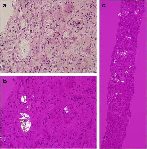
Fig. 1.

a Mild interstitial inflammation, moderate interstitial fibrosis and tubular atrophy with intratubular crystal deposition. (400X, PAS staining). b The deposits appeared strongly birefringent, forming fan-like, sheaf-like, or irregular shapes consistent with calcium oxalate crystals (400X, polarized light). c Almost all tubular lumens were filled with crystals (100X, polarized light)
