
Fig. 6.
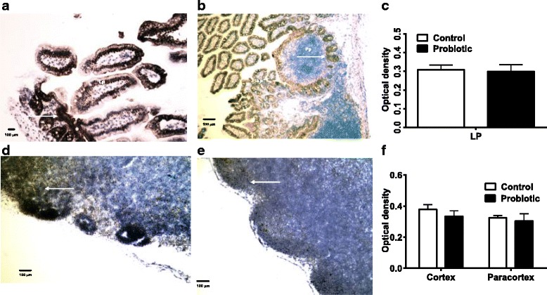
Immunohistochemical staining of RELB. The Peyer’s patches and lamina propria (a, control mice; b, probiotic-treated mice) and MLN of probiotic-treated S. enterica-infected mice (d, control mice; e, probiotic-treated mice) had RELB expressed in cells (arrows) with lymphocyte and epithelial cell morphologies. There were no significant differences in staining of lamina propria (Lp) of control or probiotic-treated mice (c). Staining in MLN from the probiotic-treated mice was not significantly different from controls, P < 0.05 by ANOVA (f)