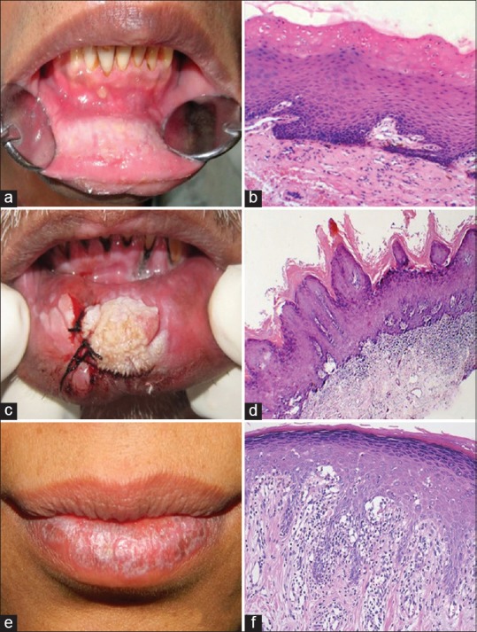
Figure 1.

(a) Oral leukoplakia: A non-scrapable, fissured gray-white lesion; (b) parakeratotic hyperkeratosis with acanthosis (Hematoxylin and eosin; 100×); (c) verrucous hyperplasia; fissured gray-white lesion; (d) hyperplastic orthokeratinized squamous epithelium thrown into verrucous folds suggestive of verrucous hyperplasia. (Hematoxylin and eosin; 40×); (e) oral Lichen Planus; White reticular striae seen on lower lip; (f) basal cell layer degeneartion with sub-epithelial inflammatory cell infiltrate suggestive of lichen planus. (Hematoxylin and eosin; 100×)
