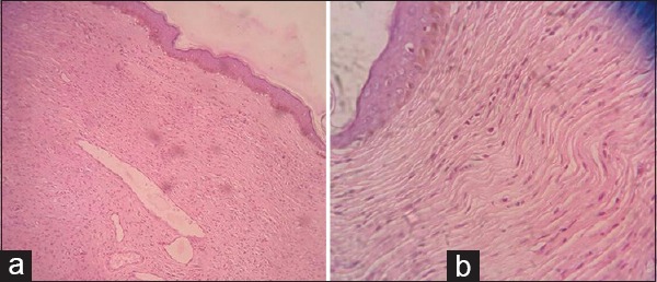
Figure 2.

(a) Epidermis revealing increased pigmentation of the basal layer due to increased proliferation of epidermal melanocytes, i.e., epidermal melanocytic hyperplasia. (Hematoxylin and eosin; ×10) (b) Dermis revealing interlacing bundles of elongated, spindle-shaped nerve cells with wavy nuclei along with intervening supportive fibrocollagenous stroma with few compressed blood vessels. (Hematoxylin and eosin; ×40)
