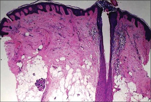
Figure 3.

Histopathology revealing partial flattening of epidermal rete ridges, perifollicular lymphocytic infiltrate, papillary dermal fibroplasias, marked reduction in the number of hair follicles, and focal replacement of follicles with early fibrotic changes (H and E, ×100)
