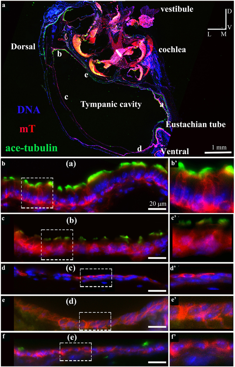
Figure 2. Dual ciliated regions in the epithelium that lines the middle ear cavity.
(a–f) A temporal bone from an adult mouse expressing the red fluorescent protein membrane Tomato protein (mT) was cryo-sectioned and staining for cilia using an antibody against acetylated α-tubulin (green) and DNA (Hoechst, blue). Ciliation was detected in both the ventral region and the dorsal region of the epithelium that lines the middle ear cavity. Panels (b) to (f) correspond to regions (a–e) in (a). (b’)–(f’) are larger views of the boxed regions in corresponding (b)–(f). The presence of cilia in the dorsal region was verified by examination of serial sections (Fig. S1). Scale: 1 mm (a) and 20 μm (b–f). The orientation designations in (a) are: L-lateral; M-medial; V-ventral; D-dorsal.

