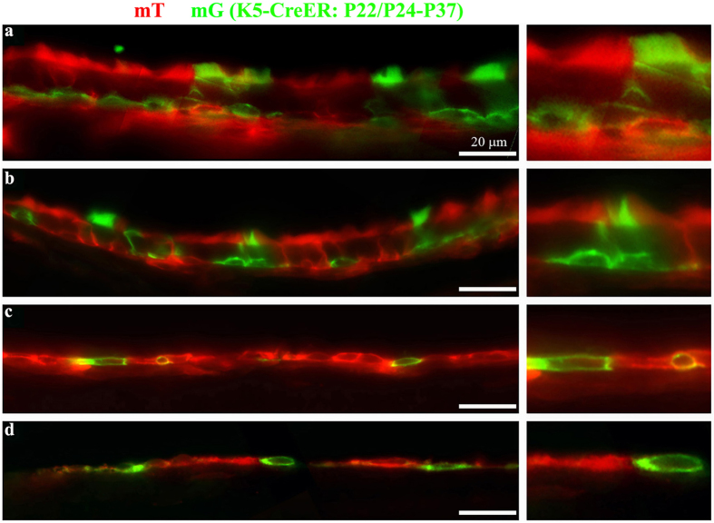
Figure 6. Keratin 5 positive cells replenish the ciliated and non-ciliated cells in the epithelium that lines the middle ear cavity.
(a–d) Mice positive for Keratin 5 (K5) Cre-ER and mT/mG transgenes were injected with Tamoxifen at postnatal days 22 (P22) and P24. The temporal bones were collected at P37 and processed for visualization of the native mT and mG signals. Panels a-d represent regions in the epithelium from the ventral region near the orifice of the Eustachian tube (a), the dorsal ciliated region (b), the non-ciliated region against the inner ear (c), and the non-ciliated region against the tympanic membrane (d). Note that the basal cells are with a flat morphology at the basal layer of the epithelium, and that some ciliated cells are green, indicating their K5-positive origin. Scale: 20 μm.

