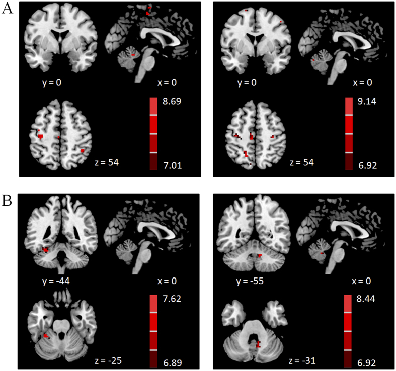
Figure 5. Brain regions more connected with the ROIs in PD patients.
Brain areas more connected with the RVM (A) or LCV (B) in PD patients than in controls during performing tapping task (left column), and dual-task (right column). Two-sample t-test, P < 0.05, FWE corrected. T-value bars are shown on the right.

