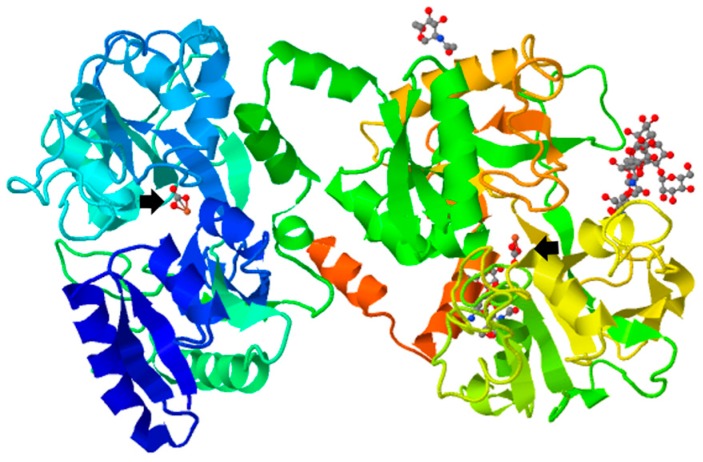
Figure 1.
Tertiary structure of bovine ferric lactoferrin. Protein Data Bank (http://www.rcsb.org/pdb/explore.do? Structure Id = 1BLF). The bovine lactoferrin is represented in rainbow ribbon diagram showing two-lobe, four-domain polypeptide. Arrows show ferric ions.

