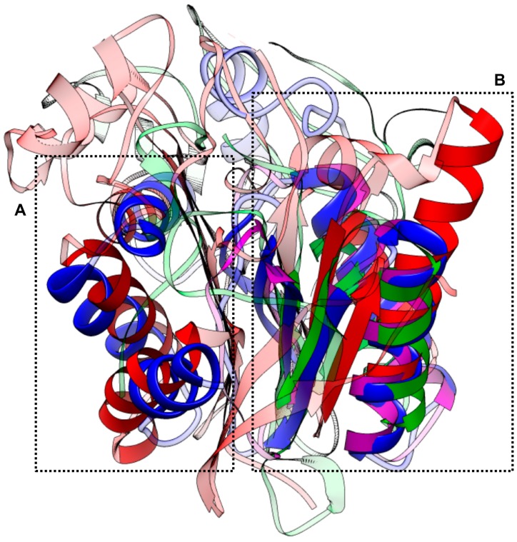
Figure 4.
The structure-based similarity and superposition. Three homologues were superposed on to CsTegue20.6 using the deconSTRUCT [40]. The superposed output is represented by the solid color, while the remainder of the structures is semi-transparent. The CsTegu20.6 (blue) was aligned to the calcium-binding protein (green, PDB ID: 1JFJ_A) on 37.1% (65/175) (A) and also both dynein light chain 1 (red, PDB ID: 1YO3_A) and dynein light chain 2 (purple, PDB ID: 1RE6_A) on 44.6% (78/175) (B).

