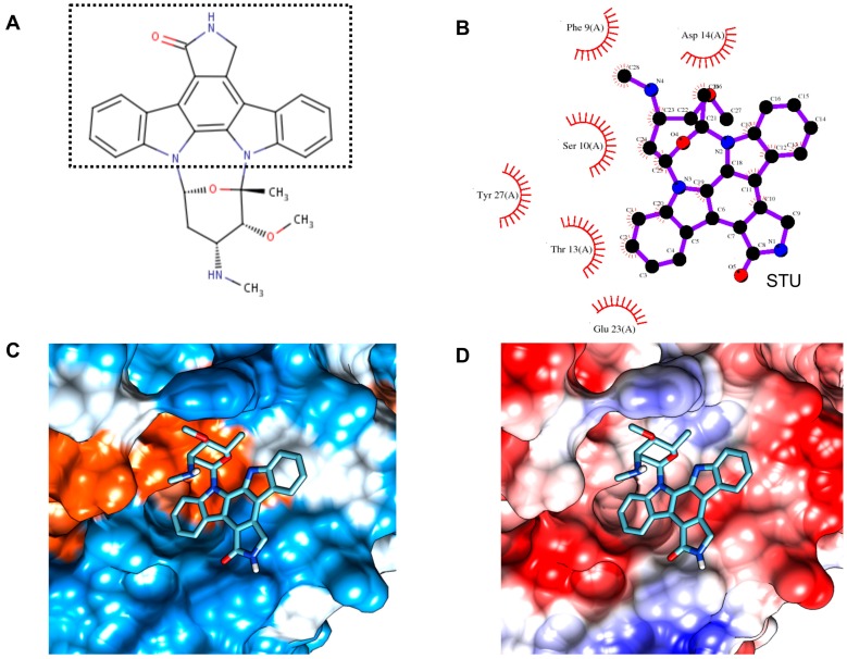
Figure 7.
Structural and detailed view of interaction of compound with CsTegu20.6. (A) STU (staurosporine). The fused indole and carbazole ring is indicated with a dotted rectangle; (B) The binding mode of STU in the CsTegu20.6 binding pocket obtained from GalaxySite [48]. Putative inhibitor and residues that are in close contact with each other are indicated in 2D diagrams. The residues, marked with red spoked arcs, involved in hydrophobic interactions with the compound; (C) Hydrophobic surface. The binding pocket is colored from blue for the hydrophilic region to orange for the hydrophobic region; (D) Electronic surface. The binding pocket is indicated from blue for the negatively-charged region to red for the positively-charged region.

