Figure 3.
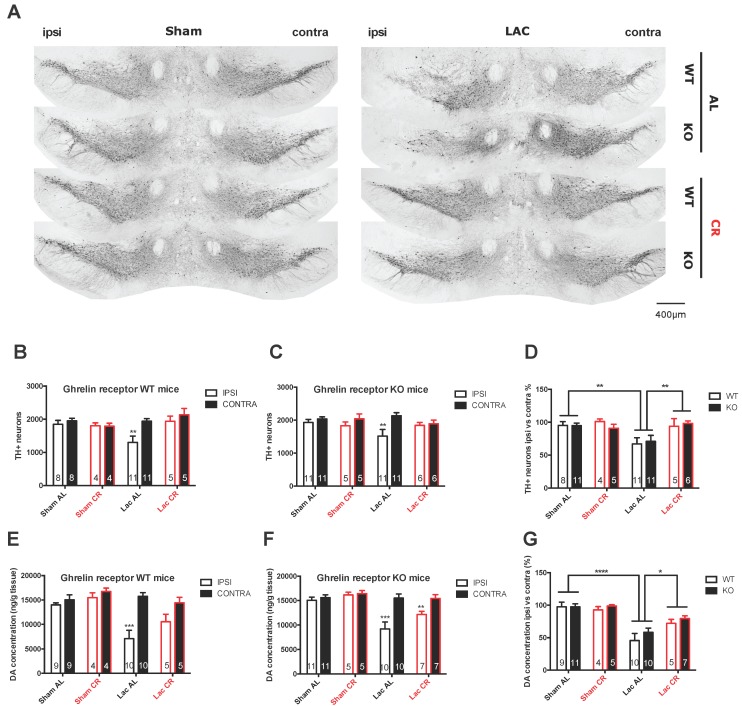
Degeneration of the nigrostriatal pathway in sham- and LAC-injected ghrelin receptor WT and KO mice fed AL or under CR. (A) Representative photomicrographs of tyrosine hydroxylase (TH) staining in the SNc of ghrelin receptor WT and KO mice under AL or CR feeding conditions seven days post-lesion (scale bar = 400 µm); (B–D) The number of TH+ neurons and percentage of TH+ cell loss of the ipsilateral SNc compared to the contralateral SNc seven days after LAC administration in CR and AL-fed WT and KO mice; ** p < 0.01 versus contra (B,C); Two-way ANOVA followed by a post-hoc Sidak’s multiple comparison test) or ** p < 0.01 versus sham AL or LAC CR (D); Two-way ANOVA followed by a post-hoc Sidak’s multiple comparisons test) (E–G); DA concentration (ng/g tissue) and percentage of DA loss in the ipsilateral striatum compared to the contralateral striatum seven days after LAC administration in CR and AL-fed ghrelin receptor WT and KO mice. *** p < 0.001 versus contra (E–F); Two-way ANOVA followed by a post-hoc Sidak’s multiple comparison test or * p < 0.01, *** p < 0.001 versus sham AL or LAC CR (G); Two-way ANOVA followed by a post-hoc Sidak’s multiple comparison test.

