Figure 2.
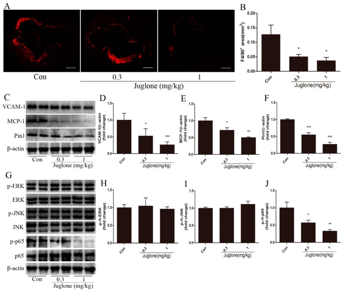
Pin1 impacts vascular inflammation and signaling pathways in the aortas of ApoE−/− mice. At ages of 18 weeks, ApoE−/− mice were sacrificed, and aortas were retrieved. The levels of macrophages accumulation in atherosclerotic lesions were examined by immunohistochemical staining of F4/80 (A) and quantified (B) (n = 7, scale bar = 50 μm); (C–F) Aortic tissues from indicated mice were subjected to Western blot for the detection of VCAM-1 (D), MCP-1 (E), and Pin1 (F) expression, normalized by levels of β-actin (n = 4), and the representative bands are shown (C); (G–J) Signal transduction in aortas was examined by Western blot: (H) for ERK, (I) for JNK, and (J) for NF-κB (n = 4), and the representative bands are shown (G). * p < 0.05, ** p < 0.01, *** p < 0.001 vs. the control group.

