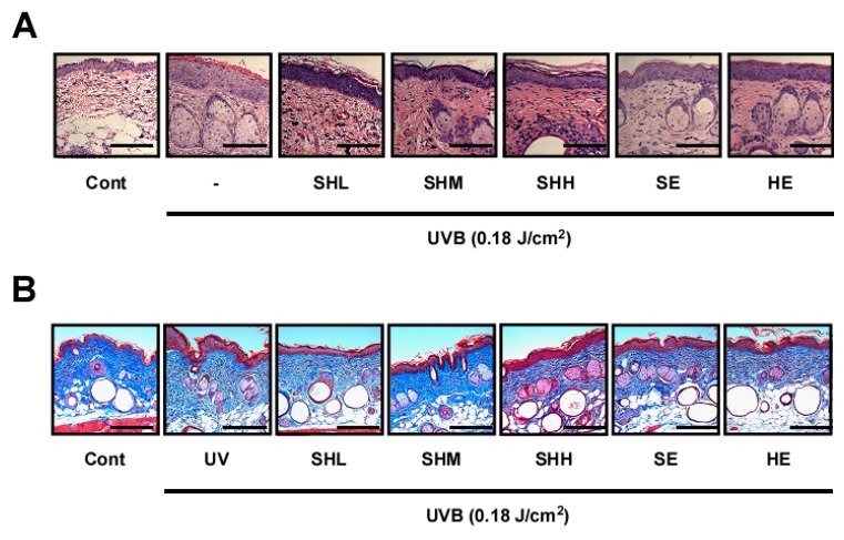
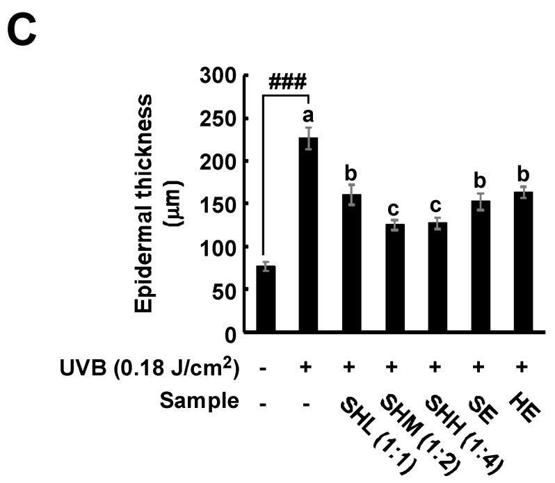
Figure 2.
Effect of SHM on UVB-induced skin inflammation and collagen degradation in hairless mice: (A,B) Dorsal skin sections were stained with hematoxylin and eosin (H&E). Scale bar is 200 μm. The epidermal thicknesses were quantified using Image J software analysis as described in the Materials and Methods. Means with letters a–c within a graph are significantly different from each other at p < 0.05. Means with letters ### within a graph are significantly different between untreated control and UVB treated group at p < 0.001. Data represent the means ± SEM (n = 5); (C) Masson’s trichrome staining for the visualization of collagen fibers as described in the Materials and Methods. Collagen fibers appear blue.


