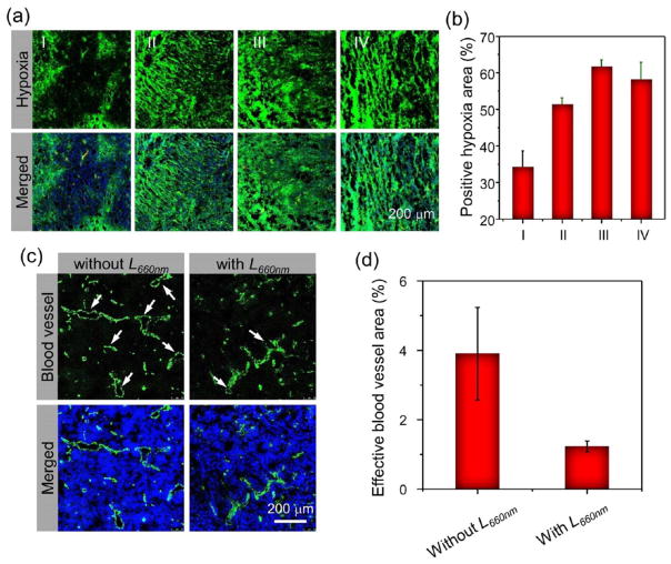
Figure 5.
Tumor hypoxia and blood vasculature evolutions induced by photodynamic treatment with AQ4N-hCe6-liposome. (a) Ex vivo immunofluorescence staining of tumor slices collected from AQ4N-hCe6-liposome injected mice with different treatments. (b) Semiquantitative analysis of the percentages of positive hypoxia regions before and after 660 nm LED light irradiation based on the images shown in (a). I, II, III, and IV stand for those tumors collected before and at 5 min, 4 h, and 24 h post-irradiation with 660 nm LED light (2 mW cm−2, 1 h), respectively. (c) Ex vivo immunofluorescence staining showing the changes of blood vessels (green) in tumors collected from the 4T1-tumor bearing mice with i.v. injection of AQ4N-hCe6-liposome before and at 4 h post-660 nm LED light irradiation (2 mW cm−2, 1 h). (d) Semiquantitative analysis of the effective blood vessel areas, which appears as opened circles indicated with white arrows, in the slices shown in (c) using the Image-Pro Plus 6.0. software.

