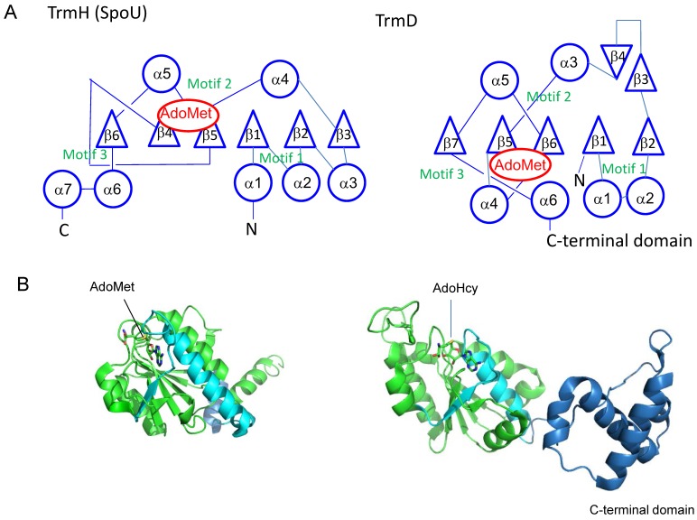
Figure 4.
(A) Topological knot structures in TrmH (SpoU) and TrmD. The representations of topologies are in accordance with the references [10,15,19]. Circles, triangles and S-adenosyl-l-methionine (AdoMet) indicate α-helices, β-strands and AdoMet binding site, respectively. (B) Cartoon models of T. thermophilus TrmH (Protein Data Bank ID: 1v2x) and Escherichia coli TrmD (Protein Data Bank ID: 1p9p) subunits. To show the knot structures, the C-terminal regions of catalytic domains are colored in cyan. The C-terminal domain of E. coli TrmD is indicated in blue. The bound AdoMet and S-adenosyl-l-homocysteine (AdoHcy) are shown by stick models.

