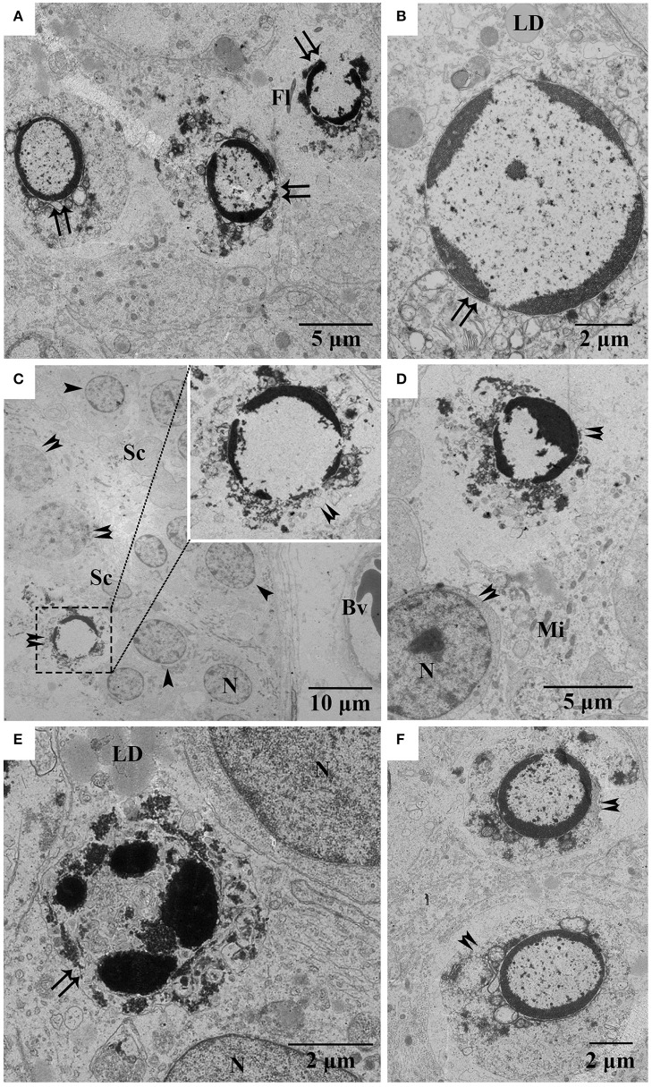
Figure 3.
Electron micrographs of various germ cell types undergoing apoptosis during spermatogenesis. (A,B) Apoptotic spermatids in April, (C,D) apoptotic spermatocytes in July, (E) apoptotic spermatids in October, (F) apoptotic spermatocytes in October. Arrowhead, spermatogonia; Double arrowhead, spermatocyte; Double arrow, spermatid; Fl, flagellum; LD, lipid droplet; Bv, blood vessel; Mi, mitochondria; N, nucleus. Scale bar = 10 μm (C), 5 μm (A,D), and 2 μm (B,E,F).

