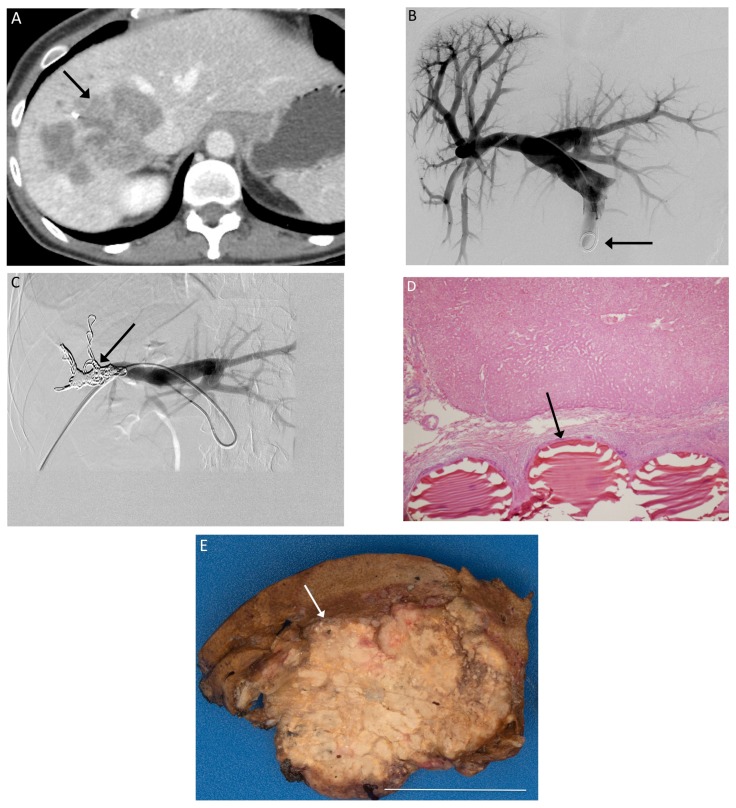
Figure 1.
(A) Pre-procedure CT imaging demonstrates a right hepatic metastasis (arrow) from colorectal cancer, with a diminutive left lobe; (B) Tranhepatic portography (arrow) is obtained after portal access is achieved via the right portal vein; (C) The right portal branches have been embolized with particles and metallic coils (arrow); (D) 200× magnification H&E stain slide demonstrates Embosphere particles within a portal vein (arrow); (E) Gross specimen after right hepatectomy demonstrates particles within the embolized right hepatic lobe. White bar indicates 5 cm.

