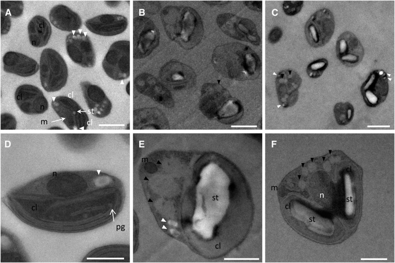
Figure 10.
Transmission electron microscopy of nutrient-replete and nutrient-depleted O. tauri cells. A and D, Control cells. Either one or duplicated chloroplasts and mitochondria could be observed, reflecting different progression stages of cells into the cell cycle. Peripheral and central arrays of thylakoids and occasionally plastoglobuli-like structures could be detected in chloroplasts. The chloroplastic starch granule, when detected, was small. Cytoplasmic arrays of granules of unknown composition were observed frequently. B and E, N-starved cells accumulated oil bodies and displayed a large starch granule in the chloroplast. Thylakoidal structures were hardly distinguishable (E). C and F, P-starved cells frequently displayed duplicated chloroplasts, each containing a large starch granule; peripheral thylakoid arrays could be detected. Black arrowheads indicate oil bodies, and white arrowheads indicate storage granules. cl, Chloroplast; m, mitochondria; n, nucleus; pg, plastoglobules; st, starch. Bars = 1 µm in general views (A–C) and 0.5 µm in individual views (D–F).

