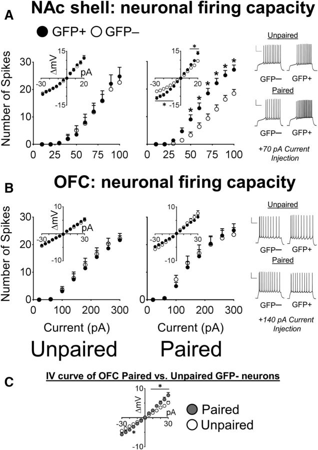
Figure 4.
GFP+ neurons activated by sucrose cues are more excitable compared with their surrounding GFP− neurons in the NAc shell but not the OFC. A, In the NAc shell, the spike counts of Paired group GFP+ neurons were significantly increased compared with the GFP− neurons after current injections (GFP−, n = 19/9; GFP+, n = 16/9). *p < 0.01. In contrast, in the Unpaired groups, the spike counts of GFP+ and GFP− neurons were similar (GFP−, n = 14/7; GFP+, n = 9/5). The I/V curve (inset) indicated that there was a large increase in the input resistance of GFP+ neurons in Paired mice, but no difference in the Unpaired mice. *p < 0.05. Example traces of GFP+ and GFP− neurons at +70 pA from the NAc shell of Paired and Unpaired mice. Scale bar, 25 mV, 250 ms. B, In the OFC, no difference in spike counts was observed between GFP+ and GFP− neurons in the Unpaired mice (GFP−, n = 12/5; GFP+, n = 14/5) and in Paired mice (GFP−, n = 11/5; GFP+, n = 17/5). The I/V curves of GFP+ and GFP− neurons in Paired and Unpaired mice are shown in the inset. Example traces of GFP+ and GFP− neurons in the OFC of Paired and Unpaired mice at +140 pA are shown. Scale bar, 25 mV, 250 ms. C, I/V curves of GFP− neurons from Paired and Unpaired mice from the OFC. *p < 0.05. Data are expressed as mean ± SEM; values to the right of GFP− and GFP+ denote number of cells recorded/number of mice used.

