Figure 2.

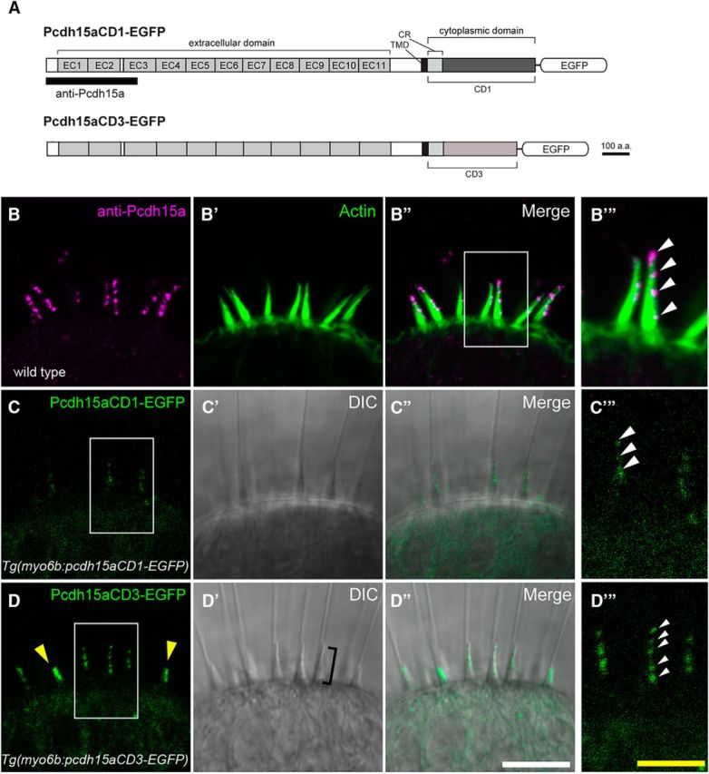
Imaging of EGFP-tagged Pcdh15a isoforms in vestibular hair cells of live fish. A, Schematic of zebrafish Pcdh15a-CD1 or Pcdh15a-CD3 fused at its C terminus to EGFP. The antigen used to generate the antibody is indicated by the black bar. B–D′′′, Representative confocal images of wild-type hair cells in the lateral cristae of inner ear at 6 dpf. B–B′′′, Pcdh15a antibody label (magenta) of the lateral crista at 6 dpf, z projection. To visualize the hair bundles, actin (green) was labeled using phalloidin (B′). C–C′′′, Image of the localization pattern of Pcdh15a-CD1-EGFP in a stable transgenic line, z projection. D–D′′′, Localization pattern of Pcdh15a-CD3-EGFP in a stable transgenic line, single section. Yellow arrowheads indicate two immature hair bundles. Bracket in D′ indicates hair bundle. B′′′, C′′′, D′′′, Higher-magnification image of area from B′′, C, and D, respectively (outlined with box). Note the staircase-like localization at the tip of the hair bundles. White scale bars in B–B′′, C–C′′, D–D′′, 10 μm. Yellow scale bars in B′′′, C′′′, D′′′, 5 μm.
