Figure 1.

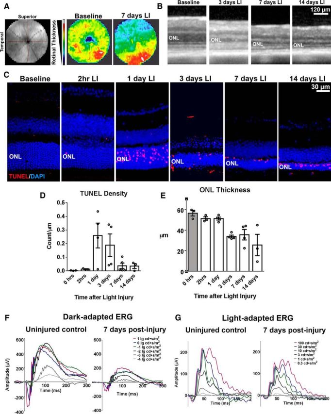
In vivo mouse model of acute light exposure-induced photoreceptor injury involves photoreceptor apoptosis, retinal atrophy, and loss of photoreceptor cell function. Young adult (2–3 months old) C57BL6J mice were dark reared for 1 week before being subjected to pupillary dilation and exposure to ambient white light at 20 × 103 lux for 2 h. The effects on retinal structure and function were evaluated at time points from 2 h to 14 d after LI. A, B, Retinal thickness and lamination was evaluated in vivo using OCT; 1.4 mm wide scan fields centered on the optic nerve were obtained. Heat maps (A) representing total retinal thickness of OCT images taken at baseline (before LI) and 7 d after LI demonstrated retinal thinning that was most marked in the superior temporal retina. Individual OCT B-scans from the superior temporal retina (B) show progressive thinning of the ONL from 3 d after LI. C–E, Histological analysis of retina in the superotemporal quadrant (1.25 mm from the optic nerve) revealed prominent emergence of apoptotic photoreceptors (as marked by TUNEL, red) in the ONL starting at 1 d after LI. Significant thinning of the ONL was observed starting at 3 d after LI. Plots in D and E show the time course of changes in the density of TUNEL-positive photoreceptors and ONL thickness after LI (column heights and error bars represent mean and SEM; n = 3–4 animals per time point). F, G, Representative ERG recordings demonstrating functional decreases at 7 d after LI relative to uninjured control mice for a- and b-wave amplitudes in dark- and light-adapted responses.
