
Figure 3.
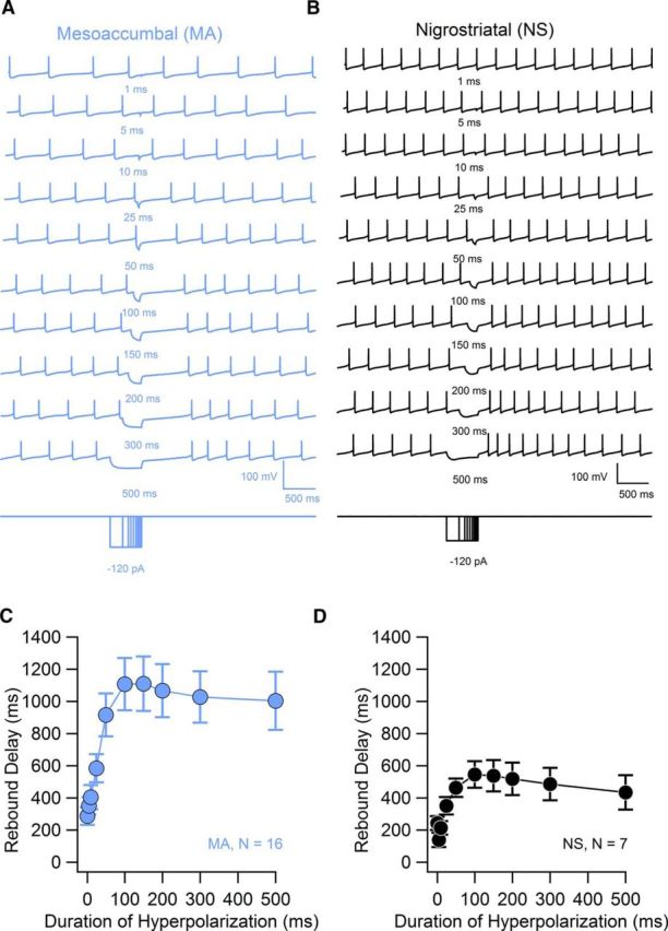
Time-dependent development of the rebound delay. A, Example of rebound pauses in a mesoaccumbal neuron in response to 120 pA hyperpolarizing current injection delivered for different durations. B, Same as in A for an example nigrostriatal neuron. C, D, Summary plots of rebound delays versus the duration of hyperpolarization in mesoaccumbal neurons (light blue symbols) and nigrostriatal neurons (black symbols). Data are plotted as averages ± SEM.