
Figure 3.
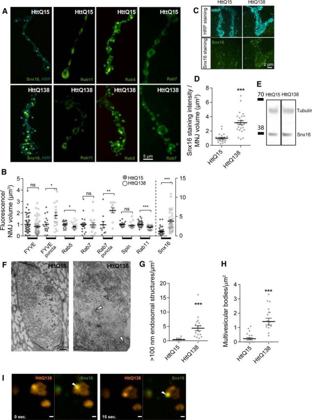
HttQ138 alter endosomal trafficking and leads to abnormal changes in a key endocytic compartments. A, Representative images of third-instar NMJ 4 showing distribution of ElavC155-driven UAS-Snx16-GFP, UAS-Rab11-GFP, UAS-Rab5-GFP, and UAS-Rab7-GFP proteins on a background of control ElavC155;UAS-HttQ15-mRFP or pathogenic ElavC155;UAS-HttQ138-mRFP lines. Anti-HRP staining was used to visualize neuronal membranes (blue). Strong accumulation of Snx16-positive compartments was observed in ElavC155;UAS-HttQ138-mRFP/UAS-Snx16-GFP larvae. The levels of UAS-Rab11-GFP and UAS-Rab5-GFP were reduced after expression of pathogenic UAS-HttQ138-mRFP (ElavC155;UAS-HttQ138-mRFP/UAS-Rab11-GFP, ElavC155;UAS-HttQ138-mRFP/UAS-Rab5-GFP and ElavC155;UAS-HttQ138-mRFP/UAS-Rab7-GFP). B, Quantification of GFP fluorescence with ElavC155-driven expression of UAS-FYVE-GFP, UAS-Rab5-GFP, UAS-Rab7-GFP, UAS-spin-GFP, UAS-Rab11-GFP, and UAS-Snx16-GFP, together with UAS-HttQ15-mRFP (filled circles) or UAS-HttQ138-mRFP (empty circles). Note the different scale (right y-axis) used for Snx16 levels (separated by dotted line). C, ElavC155;UAS-HttQ15-mRFP and ElavC155;UAS-HttQ138-mRFP lines were stained with anti-HRP (top) and anti-Snx16 (bottom). D, Quantification of Snx16-positive staining. Snx16 levels were normalized to NMJ volume determined by HRP labeling. E, Western blot analysis of Snx16 levels from head extracts of HttQ15- and HttQ138-expressing larvae. F, Representative ultramicrographs of ElavC155;UAS-HttQ15-mRFP- and ElavC155;UAS-HttQ138-mRFP-expressing boutons. Accumulation of large vacuolar structures (empty arrows) and multivesicular bodies (filled arrows) were observed in HttQ138-expressing boutons. G, Quantification of large vacuolar structures that are larger than 100 nm in diameter in HttQ15- and HttQ138-expressing larvae. H, Quantification of multivesicular bodies. Structures with inclusion of smaller vesicles inside (white filled arrow) were counted and normalized by bouton area. I, Dynamics of HttQ138 (red) and Snx16 (green) puncta in ElavC155;UAS-HttQ138-mRFP/UAS-Snx16-GFP larval NMJ boutons imaged from single confocal planes. The merged image is on the right, with colocalized puncta marked with a white arrow. Snx16 puncta colocalize with HttQ138 puncta and move together. Student's t test was used for statistical analysis. *p ≤ 0.05; **p ≤ 0.01; ***p ≤ 0.001. Error bars indicate SEM.