Figure 5.

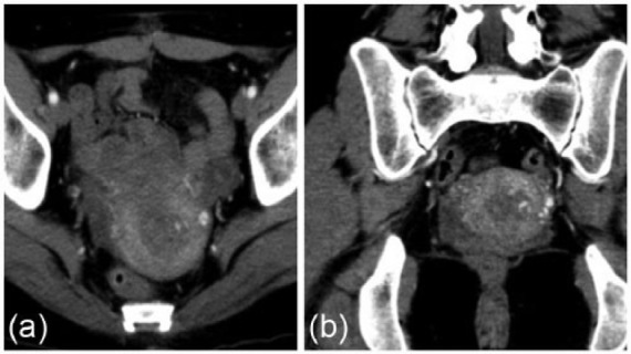
Imaging results of CT angiography in case 2. Note the hypervascular mass measuring 3 cm × 3 cm within the uterine cavity with the feeding artery being supplied by the left uterine artery. (a) Axial section (b) Coronal section.

Imaging results of CT angiography in case 2. Note the hypervascular mass measuring 3 cm × 3 cm within the uterine cavity with the feeding artery being supplied by the left uterine artery. (a) Axial section (b) Coronal section.