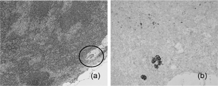
Figure 10.
(a) An example of a lymph node of a 64-year-old woman with an infiltrating ductal cell carcinoma who underwent neoadjuvant chemotherapy. The routine staining of the lymph shows a small area of suspicion for residual tumor. Please see circled portion (H&E stain ×200). (b) The presence of isolated and clusters of residual tumor cells are seen in the same lymph node upon immunostaining for cytokeratin. Please note the brown membrane staining of residual tumor cells (immunostained slide ×200).

