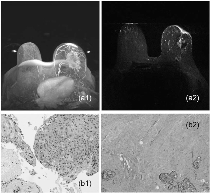
Figure 5.
(a1) An example of partial response in a 58-year-old woman with a palpable breast lesion presenting with an enhancing mass in the left breast by imaging measuring 7.9 × 5.6 × 4.7 cm3 who underwent neoadjuvant chemotherapy. (a2) Partial response is seen by a difference in the size of the lesion. (b1) This case was diagnosed as a poorly differentiated infiltrating ductal cell carcinoma (H&E stain ×400). (b2) Residual tumor cells are seen characterized by a few clusters of tumor cells in the background of fibrosis in this case (H&E stain ×200).

