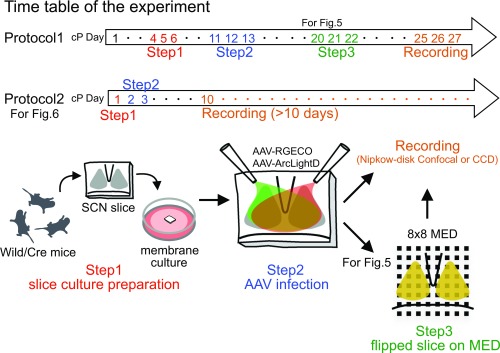
Fig. S1.
Schema of the experimental procedure. Step 1: The SCN slice was prepared from C57BL/6J or VIP/AVP-Cre newborn mice and was explanted onto a culture membrane. Step 2: Aliquots of the AAV (1 μL) harboring ArcLightD and RGECO were inoculated onto the surface of the SCN cultures. Step 3: The membrane with the cultured SCN slice was cut out, flipped over, and transferred to a glass-bottomed dish (for confocal microscopy) or a MED with 64 electrodes (for CCD imaging). Recording (confocal or CCD) was performed on days cP25–cP27 (Protocol 1) or days cP1–cP10 (Protocol 2).

