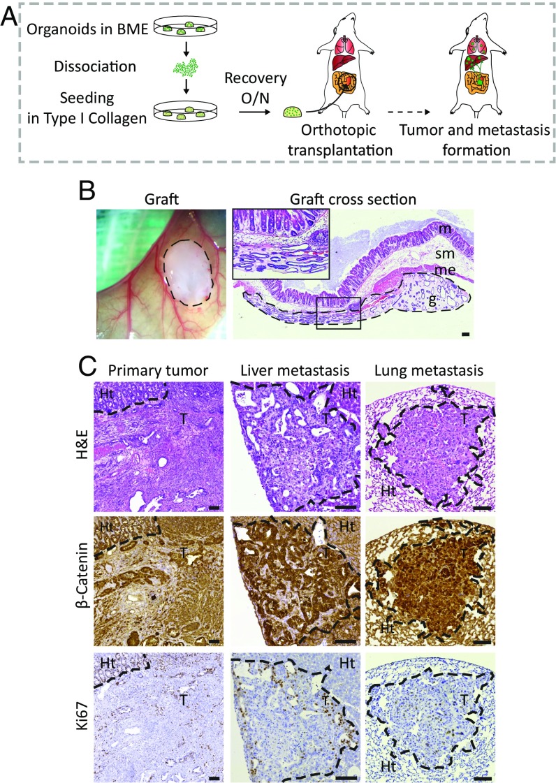
Fig. 1.
Development of an orthotopic intestinal organoid transplantation model to study CRC progression. (A) Experimental setup of the orthotopic transplantation model. The day before transplantation, 250,000 cells were plated in type I collagen. The collagen drops with the organoids were subsequently transplanted into the caecal wall of immune-deficient mice. Approximately 6–8 wk later, mice were analyzed for tumor growth and presence of metastasis. (B) Representative merged tile scan image of a transplanted collagen drop with organoids (Left) and a cross-section of the graft 1 d after transplantation (Right). g, graft; m, mucosa; me, muscolaris externa; sm, submucosa. Dashed lines highlight the graft. (C) Representative H&E, β-catenin, and Ki-67 staining of a primary tumor, and liver and long metastases. The borders between tumors tissue (T) and healthy tissue (Ht) are indicated with a dotted line. (Scale bars: 100 μm.)

