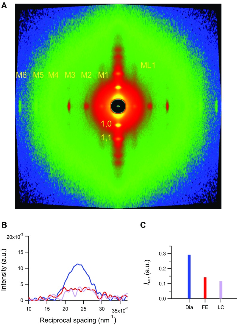
Fig. S2.
Small angle X-ray diffraction from a horizontally mounted trabecula. (A) 2D pattern collected at 1.6 m from the trabecula in diastole (Dia) showing up to the sixth order of the meridional myosin-based reflections, the first-order myosin layer line (ML1), and the equatorial reflections (1,0 and 1,1). Total exposure time was 70 ms. (B) ML1 intensity along the meridional direction in Dia (blue) and the FE (red) and LC (violet) systole. (C) ML1 integrated intensity (same color code as in B).

