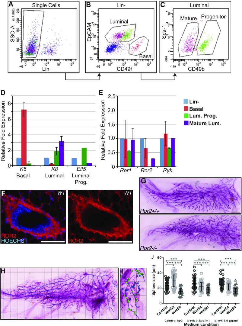
Fig. S2.
Expression of noncanonical WNT receptors in mammary epithelial populations. Mammary populations were isolated using FACS, resulting in the mammary (Linlow), basal (CD49fhi, EpCAMlow), luminal progenitor (CD49flow, EpCAMhigh,CD49bhi), and mature luminal cell (CD49flow, EpCAMhigh, CD49blow) populations (A–C). Sort purity was analyzed by performing RT-qPCR for K5, K8, and ELF5 in each of the following populations (from left to right): Lin- (blue), Basal (red), Luminal progenitors (green), and Mature Luminal (purple) (D). On these MEC populations, a subsequent RT-qPCR was performed for the putative WNT5A and WNT5B receptors Ror1, Ror2, and Ryk (n = 3). Error bars show ± SD of independently isolated, sorted, and RT-qPCR-analyzed cell population (E). Cross-sections through a cryosectioned mammary duct stained with an antibody against ROR2 and the nuclear dye Hoechst (F). Branching analysis was performed on WT and Ror2−/− outgrowths at 12 wk posttransplantation. Whole-mounts of G1 tissue display normal branching patterns (G). Shown here is a stitched image of a Ror2−/− whole-mount collected on a wide-field microscope (Keyence) at 5× resolution with the primary ductal structure outlined in blue (H). Secondary and tertiary branches were marked by green and blue boxes, respectively (I), and then branchpoints were counted. Mammosphere formation was performed in the presence of recombinant WNT5A (0.5 μg/mL) or WNT5B (0.5 μg/mL), and sphere size was measured by microscopy after 7 d (J). Sphere growth is not affected by addition of control IgG, whereas addition of anti-RYK antibody at 0.5 and 5 μg/mL lead to decrease in sphere size in the presence of WNT5A. Error bars show median measurement with interquartile range; ***P < 0.001, Student's t test.

