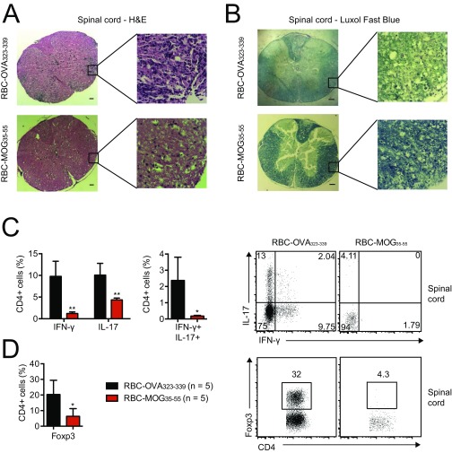
Fig. S5.
(A) H&E and (B) Luxol fast blue staining of spinal cord sections from MOG35–55-immunized mice that prophylactically received RBC-MOG35–55 or RBC-OVA323–339; these images visualize immune-cell infiltration and demyelination, respectively. (Scale bars, 100 μm.) (C) Flow cytometry analysis of CD4+ lymphocyte infiltrates into the spinal cord of diseased and protected mice at day 15–18 after immunization. (D) Frequency of Foxp3+ CD4+ regulatory T cells in the spinal cord at day 15–18 after immunization. *P < 0.05; **P < 0.01.

