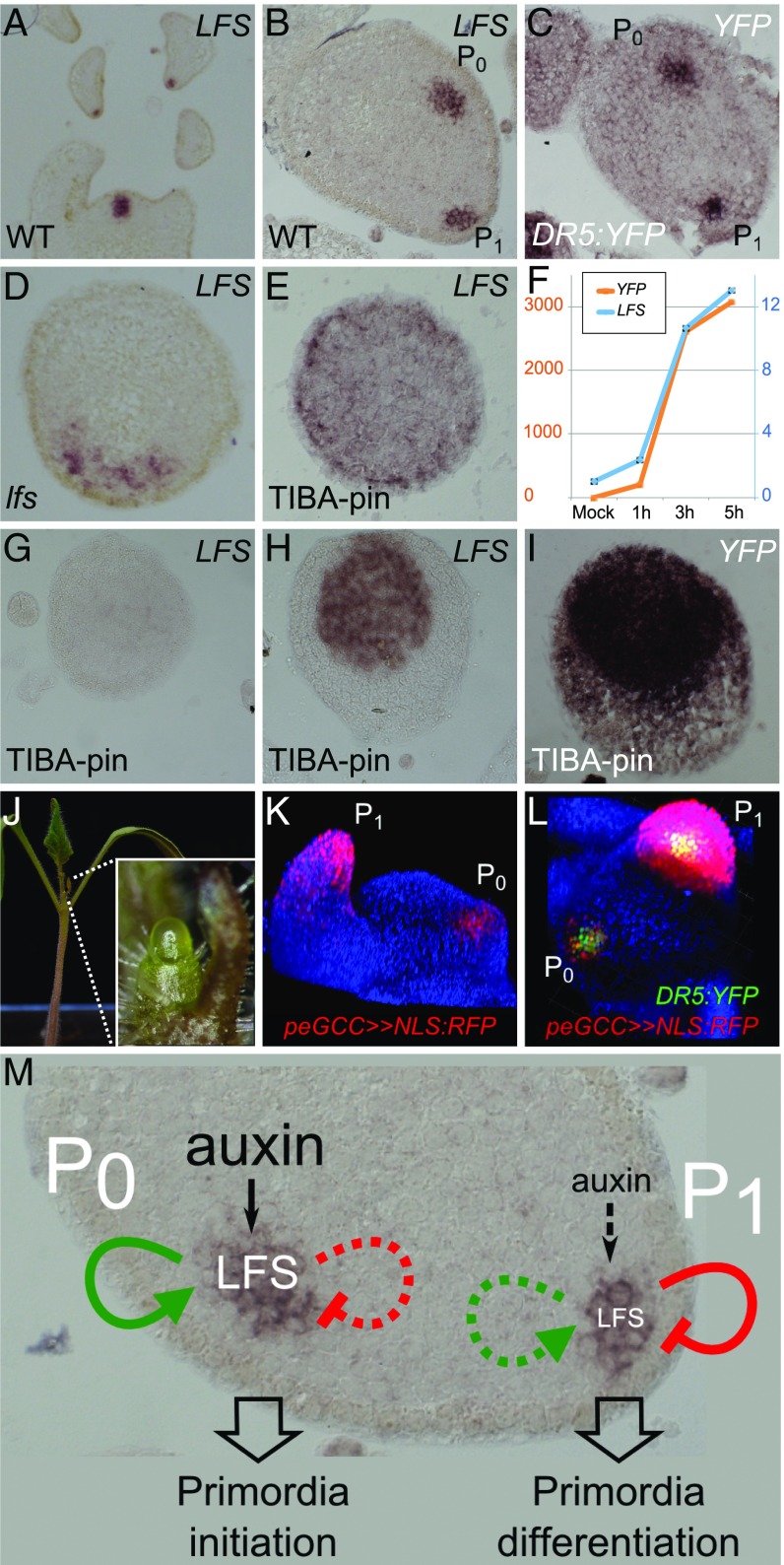
Fig. 5.
Complex regulation of LFS expression in leaf primordia. (A–E) In situ hybridization of LFS or YFP probes on WT, DR5:YFP, and lfs apices (genotypes are indicated at the Bottom Left of each panel; probes are at the Top Right). All sections were stained overnight, except E, which was stained for 4 d. (F) Relative quantification of LFS and YFP expression in response to auxin microapplication to TIBA pins. (G–I) In situ hybridization of LFS or YFP probes on DR5:YFP TIBA pins in response to microapplication of 20 mM IAA at t0 (G) and after 6 h (H and I). Probes in G–I were stained for 16 h. (J) A lfs pDR5>>LFS plant. (Inset) The pin-like apex. (K) Tomato apex with peGCC>>NLS:RFP marking young leaf primordia. (L) peGCC>>NLS:RFP DR5:YFP, viewed from the top. (M) A model for transient LFS action. Green arrows indicate GCC-dependent activation, and red arrows indicate GCC-independent inhibition.

