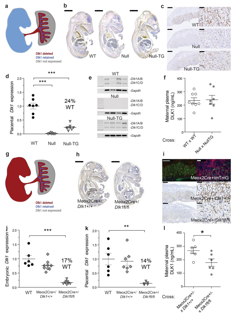
Figure 2. Fetus not placenta is the source of maternal circulating DLK1.
(a) Schematic of Dlk1 expression in Null-TG conceptuses. (b) DLK1 expression (brown staining) the WT, Null and Null–TG embryo. Scale bar 2mm. (c) DLK1 is not detected in the fetal endothelium of the Null-TG placenta but is retained in cells with large nuclei. Scale bars left 500um, right 50um. (d) Dlk1 expression in the Null-TG, WT levels, and Null placentae (n = 7–8 per group) *** p < 0.001 by Dunnett’s Multiple comparison post-test (vs WT) following One-Way ANOVA. (e) Expression of cleavable (Dlk1A/B) and membrane-bound isoforms (Dlk1C/D) of Dlk1 in WT and Null-TG placentae. (f) DLK1 levels in maternal plasma in Null x Null-TG litters (n = 7–8 females/cross). (g) Schematic of Dlk1 expression in Meox2Cre/Dlk1fl/fl conceptuses. (h) DLK1 expression in the Meox2Cre/Dlk1fl/fl embryo compared to a Meox2Cre/Dlk1+/+ control. Scale bar 2mm. (i–top) Meox2Cre crossed to the mTmG reporter results in GFP+ cells following Cre excision, and mTomato+ in non–recombined cells. (i–bottom) Dlk1 is not detected the fetal endothelium of the Meox2Cre/Dlk1fl/fl placenta but some labyrinthine expression of DLK1 is retained. Scale bars left 500um, right 50um. (j) and (k) Dlk1 expression in the WT, Meox2Cre/Dlk1+/+ and Meox2Cre/Dlk1fl/fl embryo and placenta (5–8 per group), **p < 0.01, ***p < 0.001 as in (d). (l) Maternal plasma DLK1 in Meox2Cre+/- females crossed with Dlk1+/+ (control) or Dlk1fl/fl males (n = 6 females/cross), *p < 0.05 compared by Students’ t–test. All measurements performed at E15.5. Vertical bars show mean ± s. e. m.

