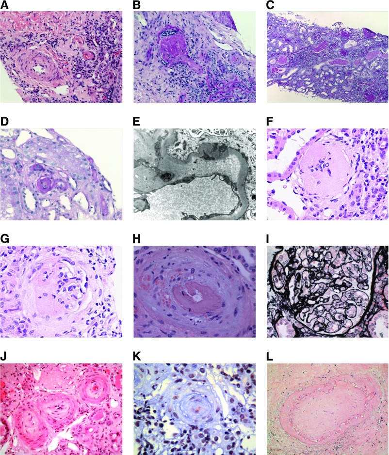
Figure 2.
Thrombotic microangiopathy in renal biopsy from patients with INF2 mutations. Renal biopsies. Native renal biopsy from family 1, patient III:2. (A) Two arterioles (right) showing features of active thrombosis and a small artery (left) with relatively slight intimal edema with fibrosis (hematoxylin and eosin stain [H&E]). (B) Glomerulus (left) showing global sclerosis and occluded arteriole (right) (periodic acid–Schiff [PAS]). Native renal biopsy from family 1, patient II:2 demonstrating end-stage changes with diffuse global sclerosis (C). Renal transplant biopsy from family 1, patient II:2 demonstrating (D) an occluded arteriole (PAS) and (E) a capillary loop with abundant subendothelial fluffy material (electron microscopy). Native renal biopsy from family 2, patient III:2 demonstrating (F) a sclerosed glomeruli and (G) a segmental sclerosing lesion. Renal transplant biopsy from family 2, patient III:1. Mucoid intimal thickening is seen in an interlobular artery with red cell fragmentation in the wall and luminal thrombus (H). Mesangiolysis is also seen 3–6 o’clock (I) (silver stain). Native renal biopsy from family 2, patient III:2 showing a subacute/chronic arterial TMA with fibroproliferative obliteration of small arteries and arterioles (J) (H&E) and (K) (trichrome). Renal transplant biopsy from family 2, patient III:2 demonstrating end stage change with fibrous obliteration of arteries. (L) (H&E).

